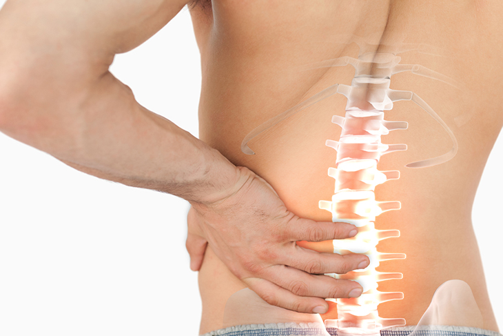
Anatomy and physiology of the sacroiliac joints
The sacroiliac joint, abbreviated as “SI” joint, is a connection of two bones just below the lumbar vertebrae (your lower back). This joint is composed of the sacrum and ilium bones. Just as the keystone in a masonry arch serves to maintain the structural integrity of doorways and ceilings, the sacrum is a biological equivalent to the structural integrity of the pelvis.
There are 2 parts to the SI joint; on either side of the sacrum we have 2 iliums (place your hands on your ‘hips’ and you’re feeling the top of the ilium) and between the placements of your hands being on your hips lays the sacrum. This is the “SI joint”. Previous school of thought believed this joint to be relatively ‘fixed’, or extremely stable. However, more up-to-date research outlines how the mobility and synchronicity of movement at this joint plays an extremely important role in the normal motion of the human body.
Normal movement of the sacrum in relation to the ilium is described as ‘nutation’ (and conversely, ‘counter nutation’), which can be defined as oscillatory movement of the axis of a rotating body1. Another way to think of this is to imagine the top of the sacrum moving forward and down compared to the bottom, and then the opposite would then be counter nutation. These motions occur in conjunction with other movements like walking, bending forward/backward, and even breathing!


